The brain as a central computer is a critical component of animal central nervous system. Melatonin, an endogenous neurohormone, predominantly synthesized and secreted by the pineal gland participates in several important physiological functions due to its high lipid and water solubility.The aim of this study was to explore whether melatonin regulates the brain cell apoptosis caused by carbon ions in mice at the level of signal transduction pathway.
Researchers in Institute of Modern Physics, Chinese Academy of Sciences (IMP) investigated the protective mechanisms of melatonin on the brain injury caused by carbon ion beams at the initial energy of 270 MeV/u. They identified the relationships between melatonin and the mitochondria mediated endogenous signal pathway involved the apoptosis of nerve cells and the activation of transcription factor Nrf2.
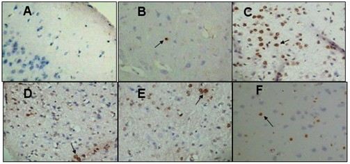
The result shows that melatonin supplementation was better able to prevent mitochondria-mediated apoptotic pathway and mitigate apoptotic rate (Fig.1) through its two functions: 1), Melatonin treatment maintained mitochondrial membrane potential as well as regulated the opening of mitochondria permeability transition to preserve mitochondrial structural integrity and function via attenuating ROS production. Moreover, treatment with melatonin diminished cytochrome c release from mitochondria, down-regulated Bax/Bcl-2 ratio and caspase-3 levels. 2), This publication is the first report showed that administration of melatonin pronouncedly elevated the expression of Nrf2 to induce antioxidant enzyme activity indirectly (Fig.2), consequently abolished carbon ion–induced brain injury.
This research has been published on J Pineal Res.2012,52(1):47-56.
Web Link: http://onlinelibrary.wiley.com/doi/10.1111/j.1600-079X.2011.00917.x/full

Fig.1 TUNEL stained histology of mouse brain sections at 12 hr after carbon ion irradiation with or without melatonin. (Original magnification 200×)
A: negative control group; B: control group; C: IR group; D: 1 mg/kgMel + IR group; E: 5 mg/kgMel + IR group; and F: 10 mg/kgMel + IR group . IR: Irradiation,Mel: Melatonin. (Image by IMP)

Fig.2 Effect of melatonin on the expression of Nrf2 protein in carbon ion–irradiated mouse brain.
Note: +: positive treatment, -: negative treatment. IR: Irradiation,Mel: Melatonin.(Image by IMP)

